
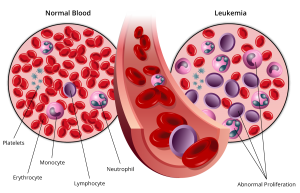
Leukemia, also known as blood cancer, is a kind of clonal malignant disease with abnormal hematopoietic stem cells (HSC). The pre-leukemic HSC clones lose the ability to further differentiate and mature, thus stagnate at different stages of cell development. It usually begins in the bone marrow and result in high numbers of abnormal blood cells. A large number of leukemia cells proliferate and spill over into the bloodstream and spread to other organs of the body, at the same time, inhibit normal hematopoiesis. Symptoms may include anemia, bleeding, infection and infiltration of various organs.
Classification of Leukemia
Leukemia is classified in two ways. First, the disease can be either chronic (slow-growing) or acute (more aggressive). Second, leukemia is classified based on the types of leukemia cells present. This is determined by where the disease started.
| Cell type | ||
|
Myeloid |
Lymphoblastic |
|
|
Acute |
Acute myelogenous leukemia (AML)
|
Acute lymphoblastic leukemia (ALL)
|
|
Chronic |
Chronic myelogenous leukemia (CML)
|
Chronic lymphocytic leukemia (CLL)
|
The cure rate and survival time of leukemia patients have a great relationship with the type of leukemia. It has been found clinically that different types of leukemia have different therapeutic effects. Without intervention, the natural course of the acute leukemia is several months to half a year, the natural course of the chronic leukemia is about five years. Acute and chronic leukemia basically do not affect life expectancy after treatment.
With the development of modern medicine, molecular biology and biogenetics, the discovery and use of new treatments and new drugs have greatly improved the prognosis of leukemia. Systematic diagnosis and treatment can make most leukemia patients survive disease-free for a long time, and even recover.
The causes of Leukemia
There is no single known cause for any of the different types of leukemias. The different leukemias likely have different causes.
1.Virus
Scientists have done experiments on mice, cats, cows and other animals, and isolated leukemia virus from their spontaneous leukemia virus. The viral etiology of human leukemia has been studied for decades, but so far only adult T-cell leukemia is caused by HTLV-1 virus. HTLV-1 is understood to be transmitted primarily through blood transfusion or b breastfeeding.
2.Radiation
One large dose of radiation or multiple small doses of radiation can possibly lead to leukemia. Whole body irradiation, especially bone marrow irradiation, can lead to a decrease in the activity of blood cell precursors in the bone marrow, as well as a decrease in immunity, making it susceptible to infection by bacteria, viruses, and fungi. Several months after irradiation, chromosomal breaks and aberrations can still be observed, and then acute nonlymphocytic leukemia, acute lymphocytic leukemia, and chronic myeloid leukemia can be induced. There is no evidence for whether diagnostic radiation can cause leukemia, but intrauterine radiation of pregnant women can increase the risk of leukemia in babies after birth.
3.Chemicals
The pathogenic effect of benzene is relatively certain, and the types of leukemias it caused are mainly AML, CML, and erythroleukemia, a rare form of AML. Alkylating agents and cytotoxic drugs can cause secondary leukemia. Secondary leukemia caused by previous exposure to radiation or chemotherapy exposure for another cancer,it is mainly acute non-lymphocytic leukemia, and there is often a period of pancytopenia before the onset.
4.Genetic factors
Leukemia is generally not considered a hereditary disease. However, having a close family member with leukemia increases your risk of leukemia. Among the leukemia patients, those with a family history of leukemia accounted for 8.1%, while only 0.5% in the control group. The incidence of ALL in consanguineous marriages was 30 times higher than expected. Monozygotic twins, if one person is suffering from leukemia, the other has a 20% chance of developing leukemia. A small number of leukemia are familial, and familial leukemia accounts for 7% of the total number of leukemia cases.
Blood test: can determine if you have abnormal levels of red or white blood cells or platelets— which may suggest leukemia.
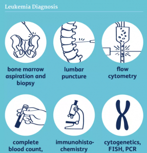
Bone marrow aspiration: a procedure to remove a sample of bone marrow from your hipbone. The fluid sample gets tested in a lab for leukemia cells. A bone marrow biopsy helps determine the percentage of abnormal cells in your bone marrow, confirming a leukemia diagnosis.
Cytochemistry: looks at how the cells in the bone marrow take up certain stains, and it can be helpful in distinguishing ALL from AML. Tests can include both flow cytometry and immunohistochemistry.
Chromosome Studies: Leukemia cells very often have changes in the chromosomes in the DNA of each cell. Cytogenetics involves microscopic examination of the chromosomes of cancer cells.
Gene Studies: Fluorescent In Situ Hybridization (FISH) and PCR can find changes in chromosomes and genes that can’t be identified through cytogenetics. PCR is very sensitive for finding the BCR/ABL gene, even when other signs of CML aren’t found on chromosome testing. PCR can also be used to determine the amount of minimal residual disease (MRD), the small amount of cancer cells left in the body after treatment. This testing can be done on a bone marrow or a blood sample.
Imaging: Imaging tests are not usually used as a diagnostic method for leukemia, as blood-related cancers like leukemia don’t often form tumors. It may be helpful, however, in staging some leukemias, such as CLL.
Leukemia patients are often accompanied by chromosomal abnormalities. Abnormal genes formed by chromosomal recombination are called fusion genes.
More than half of leukemia lack cytogenetic changes, and each leukemia cell is the result of accumulation of multiple gene mutations. A comprehensive understanding of leukemia mutations at genetic level is helpful for accurate diagnosis and classification, as well as the assessment of prognosis and formulation of individualized treatment plans. Gene mutation screening is the most effective method.
Early diagnosis and prognosis
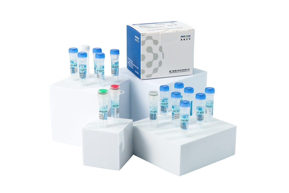
Leukemia Fusion Genes (Q30) Screening Kit is a qualitative in vitro diagnostic test for screening of 30 fusion genes resulted from chromosome translocations involved in chronic and acute leukemia. All the 30 fusion genes include more than 155 clinically relevant chromosomal breakpoints.
Leukemia Fusion Genes (Q51) Screening Kit is a qualitative in vitro diagnostic test for screening of 52 fusion genes, which include more than 182 clinically relevant chromosomal breakpoints
Both kits can better facilitate accurate diagnostic typing and prognosis of leukemia.
During treatment and complete remission
Zeesan offers 53 kinds of individual leukemia fusion gene quantitative kit for the monitoring of treatment effects, which provide a reference for clinical adjustment of treatment plans and strategies.
Jul 24,2023